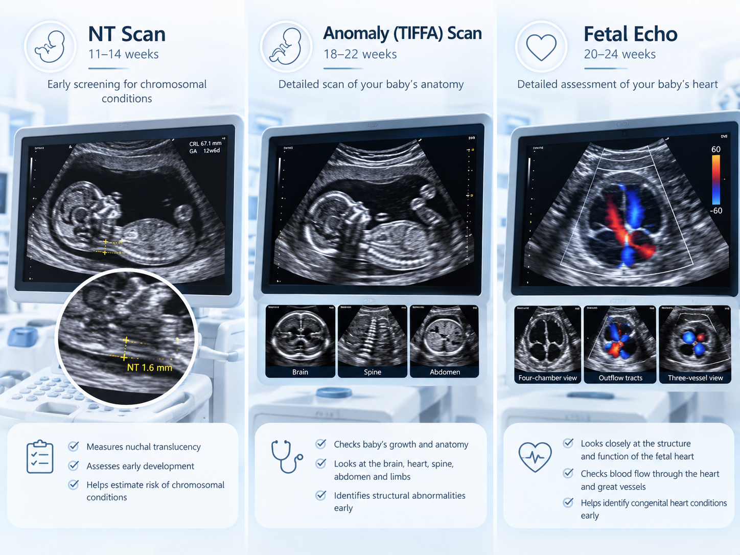
Pregnancy involves several important ultrasound scans, and many parents become confused about the difference between an NT scan, anomaly scan, and fetal echo.
A common question is: Should I get an NT scan or anomaly scan, and is fetal echo another version of the same test?
The answer is no. Each scan has a different purpose and usually takes place at a different stage of pregnancy.
In simple terms:
- NT scan mainly assesses early fetal development and markers associated with chromosomal abnormalities.
- Anomaly scan or TIFFA scan checks the baby’s anatomy in greater detail.
- Fetal echocardiography focuses specifically on the structure and function of the baby’s heart.
At Paaranu IVF, fetal medicine care is led by Dr. Jayesh Patel, MD, D.G.O., Clinical Director, who has more than 25 years of experience in gynecology, fertility, and fetal medicine.
What Is the Difference Between NT Scan, Anomaly Scan, and Fetal Echo?
These three scans are not interchangeable.
| Scan | Usual Timing | Main Purpose |
|---|---|---|
| NT Scan | Around 11–14 weeks | Early fetal assessment and nuchal translucency measurement |
| Anomaly/TIFFA Scan | Around 18–22 weeks | Detailed examination of fetal anatomy |
| Fetal Echo | Commonly around 18–22 weeks when indicated | Detailed assessment of the baby’s heart |
ISUOG guidance places the first-trimester ultrasound around 11 to 14 weeks, while the routine mid-trimester anomaly scan generally occurs around 18 to 22 weeks. Fetal echocardiography is also commonly performed in the second trimester when a detailed heart evaluation is indicated.
What Is an NT Scan?
NT stands for Nuchal Translucency.
The scan measures the fluid-filled space at the back of the baby’s neck during early pregnancy.
A certain amount of fluid is normal. However, an increased NT measurement can be associated with a higher chance of:
- Chromosomal abnormalities
- Certain structural abnormalities
- Congenital heart defects
- Some genetic syndromes
The NT scan is a screening test, not a diagnostic test.
An abnormal or increased NT result does not automatically mean that the baby has a medical condition.
When Is an NT Scan Done?
The NT assessment is generally performed between approximately 11 weeks and 13 weeks + 6 days, when the crown-rump length falls within the appropriate range for reliable measurement.
The wider first-trimester anatomical scan window may extend to around 14 weeks depending on the guideline and clinical setting.
Because NT measurement has a specific timing window, parents should avoid delaying the scan beyond the date recommended by their obstetrician or fetal medicine specialist.
What Does the NT Scan Check?
An NT scan may assess:
- Nuchal translucency thickness
- Crown-rump length
- Fetal heartbeat
- Early fetal anatomy
- Skull and brain appearance
- Arms and legs
- Stomach and bladder
- Placental location
- Number of babies
- Chorionicity in twin pregnancies
Modern first-trimester ultrasound should not focus only on the NT measurement. ISUOG recommends a broader early anatomical assessment because some major structural abnormalities can already appear during the 11–14-week scan.
Does an NT Scan Diagnose Down Syndrome?
No.
An NT scan can contribute to risk assessment for chromosomal abnormalities such as trisomy 21, commonly known as Down syndrome.
Doctors may combine the NT measurement with:
- Maternal age
- Blood tests
- Nasal bone assessment
- Other ultrasound markers
- NIPT or cell-free DNA testing
If screening suggests a higher risk, your doctor may discuss further tests.
These may include additional genetic screening or diagnostic procedures depending on the individual pregnancy.
What Does Increased NT Mean?
An increased NT measurement means the fluid space behind the baby’s neck appears thicker than expected.
It can be associated with:
- Chromosomal abnormalities
- Congenital heart disease
- Genetic conditions
- Structural abnormalities
However, some babies with increased NT have normal genetic testing and develop normally.
ISUOG guidance states that an NT measurement of 3.5 mm or more is an indication for detailed cardiac evaluation because of the increased association with congenital heart disease.
Your fetal medicine specialist may therefore recommend additional testing and a fetal echo if NT is significantly increased.
What Is an Anomaly Scan?
The anomaly scan is a detailed ultrasound examination performed during the second trimester.
It is also commonly called:
- Anatomy scan
- Level II scan
- Target scan
- TIFFA scan
TIFFA means Targeted Imaging for Fetal Anomalies.
The purpose is to examine the developing baby’s organs and structures systematically.
ISUOG describes the mid-trimester scan as an important routine ultrasound for detecting major fetal structural abnormalities.
When Is the Anomaly or TIFFA Scan Done?
The detailed anomaly scan is commonly performed between approximately 18 and 22 weeks of pregnancy.
At this stage, the baby is usually large enough for the examiner to assess individual structures in greater detail.
The exact appointment may depend on:
- Gestational age
- Fetal position
- Multiple pregnancy
- Previous scan findings
- Maternal medical history
- Local fetal medicine protocol
What Does the Anomaly Scan Check?
The anomaly scan may examine:
- Brain
- Skull
- Face
- Spine
- Heart
- Chest
- Stomach
- Kidneys
- Bladder
- Abdominal wall
- Arms and legs
- Placenta
- Umbilical cord
- Amniotic fluid
- Fetal growth
The fetal heart forms part of the routine anatomy scan, but this cardiac screening is not always the same as a full fetal echocardiogram.
Is the Anomaly Scan the Same as TIFFA Scan?
In everyday clinical use, TIFFA scan and anomaly scan usually refer to the detailed mid-pregnancy anatomical examination.
Both aim to evaluate the fetus for structural abnormalities.
Different hospitals may use terms such as:
- TIFFA
- Level II ultrasound
- Target scan
- Anatomy scan
- Anomaly scan
The terminology can differ, but the overall purpose is a detailed structural evaluation of the baby.
What Is the Difference Between NT Scan and TIFFA Scan?
Parents often search TIFFA vs NT scan, but these tests occur at different stages and answer different questions.
NT Scan
The NT scan occurs in the first trimester and focuses on early anatomy plus chromosomal-risk markers such as nuchal translucency.
TIFFA or Anomaly Scan
The TIFFA scan occurs in the second trimester and provides a much more detailed assessment of the baby’s organs and structural development.
Therefore, a normal NT scan does not replace the anomaly scan.
Even if the first-trimester scan appears reassuring, the second-trimester anatomical scan remains important because some structural abnormalities become easier to identify later.
What Is a Fetal Echo?
A fetal echocardiogram, or fetal echo, is a specialised ultrasound examination focused specifically on the baby’s heart.
It provides a more detailed cardiac assessment than the routine heart views included in the anomaly scan.
A fetal echo may assess:
- Four chambers of the heart
- Heart valves
- Great arteries
- Blood flow
- Heart rhythm
- Connections between major vessels
- Cardiac function
The goal is to identify or clarify congenital heart abnormalities before birth.
When Is a Fetal Echo Usually Done?
A comprehensive fetal echocardiogram is commonly performed around 18 to 22 weeks of pregnancy, although specialists may perform earlier or later examinations in selected situations.
If an early scan raises a significant concern, a specialist may assess the fetal heart earlier and repeat the examination later as the heart develops further.
Does Every Pregnant Woman Need a Fetal Echo?
Not necessarily.
Every routine anomaly scan should include basic cardiac assessment, but a full fetal echocardiogram is usually reserved for pregnancies with a specific indication or increased risk.
Your obstetrician or fetal medicine specialist will decide whether you need one.
Who May Need a Fetal Echo?
Doctors may recommend fetal echocardiography when there is:
- A suspected heart abnormality on routine ultrasound
- Increased NT
- Family history of congenital heart disease
- Certain fetal genetic abnormalities
- Abnormal fetal heart rhythm
- Another fetal structural abnormality
- Pregestational diabetes
- Certain maternal autoimmune antibodies
- Monochorionic twins
- Selected medication or teratogen exposures
Current ISUOG fetal cardiac screening guidance lists these among recognised indications for detailed fetal echocardiography.
IVF or ICSI conception may also lead to consideration of fetal echo depending on the clinical situation and local screening practices.
Why Is Increased NT Linked to Fetal Echo?
An increased NT measurement has an association with congenital heart abnormalities, even when chromosome testing is normal.
ISUOG specifically identifies NT ≥3.5 mm as an indication for detailed cardiac evaluation.
Therefore, a pregnancy with increased NT may require:
- Genetic counselling or testing
- Detailed anomaly assessment
- Fetal echocardiography
- Additional follow-up scans
The exact pathway depends on the NT measurement and any other ultrasound findings.
Can a Normal Anomaly Scan Replace a Fetal Echo?
Not always.
The anomaly scan assesses the entire fetus and includes screening views of the heart.
A fetal echo performs a more specialised assessment of cardiac anatomy and function.
If there are no risk factors and cardiac views during the anomaly scan appear normal, a fetal echo may not be necessary.
However, when specific indications exist, a normal routine scan does not automatically remove the need for specialist fetal echocardiography.
Can a Fetal Echo Replace the Anomaly Scan?
No.
A fetal echo focuses on the heart.
The anomaly scan checks the:
- Brain
- Face
- Spine
- Chest
- Abdomen
- Kidneys
- Limbs
- Placenta
- Amniotic fluid
- Heart and other organs
Therefore, fetal echo complements rather than replaces the routine anomaly scan.
Do You Need All Three Scans?
Many pregnancies will have:
- A first-trimester ultrasound/NT assessment
- A mid-trimester anomaly scan
A fetal echo is added when the obstetrician or fetal medicine specialist identifies a reason for more detailed cardiac assessment.
So the scans are not simply three versions of the same ultrasound.
Each has a different role.
What Happens if the NT Scan Is Normal?
A normal NT result is reassuring, but it does not rule out every chromosomal or structural abnormality.
You should still attend your recommended anomaly scan later in pregnancy.
Even highly effective genetic screening cannot detect every structural abnormality, which is why ultrasound remains an important part of pregnancy care.
What Happens if the Anomaly Scan Shows a Problem?
If the anomaly scan identifies or suspects an abnormality, your doctor may recommend:
- Repeat targeted ultrasound
- Fetal medicine consultation
- Fetal echocardiography
- Genetic counselling
- NIPT or additional screening
- Amniocentesis in selected situations
- Follow-up growth scans
The next step depends on which organ appears affected and how certain the ultrasound finding is.
What Happens if a Heart Problem Is Seen?
If the routine scan suggests a congenital heart defect, a specialist fetal echocardiogram should assess the baby’s heart in greater detail.
Fetal cardiac specialists may evaluate:
- Exact anatomy
- Severity of the defect
- Heart function
- Associated abnormalities
- Possible genetic associations
- Expected care after delivery
Early diagnosis can also help the obstetric and neonatal teams plan the safest place and timing of delivery when specialised newborn cardiac care may be required.
Are NT, Anomaly Scan and Fetal Echo Safe?
These tests use diagnostic ultrasound and do not use ionising radiation.
When appropriately performed for medical reasons by trained professionals, ultrasound forms a standard part of prenatal assessment.
The number and timing of scans should follow your individual pregnancy needs rather than unnecessary repeated scanning.
Where Can You Get Fetal Medicine Scans in Surat?
Paaranu IVF provides fetal medicine services in Surat, including:
- NT scans
- Detailed anomaly/TIFFA scans
- Fetal echocardiography
- Doppler studies
- High-risk pregnancy assessment
- Genetic counselling and testing
The clinic states that fetal medicine services are led by Dr. Jayesh Patel, MD, D.G.O., who has over 25 years of experience across gynecology, fertility and fetal medicine.
For a detailed cardiac assessment, you can learn more about Fetal Echo in Surat.
You can also explore Paaranu IVF’s Fetal Medicine Specialist in Surat service for NT, anomaly, Doppler, and high-risk pregnancy monitoring.
Which Scan Should You Book and When?
A simple pregnancy scan timeline looks like this:
Around 11–14 Weeks
NT / first-trimester scan
Purpose:
- Early anatomy
- Nuchal translucency
- Chromosomal risk assessment
- Early structural evaluation
Around 18–22 Weeks
Anomaly / TIFFA scan
Purpose:
- Detailed assessment of fetal anatomy
- Growth
- Placenta
- Amniotic fluid
- Screening of the fetal heart
Around 18–22 Weeks When Indicated
Fetal echocardiography
Purpose:
- Detailed evaluation of fetal heart structure, rhythm, blood flow, and function
Your doctor may adjust this schedule if the pregnancy is high-risk or if an earlier scan identifies a concern.
Frequently Asked Questions
Which is more important: NT scan or anomaly scan?
Both are important because they assess the baby at different stages. The NT scan focuses on early development and chromosomal-risk markers, while the anomaly scan performs a much more detailed structural examination later in pregnancy.
Is TIFFA the same as an anomaly scan?
Yes, the terms are commonly used for the detailed mid-pregnancy anatomical scan performed around 18–22 weeks.
If NT is normal, do I still need an anomaly scan?
Yes. A normal NT scan does not rule out every structural abnormality, so the routine second-trimester anomaly scan remains important.
If anomaly scan is normal, do I need fetal echo?
Not everyone does. Fetal echo is usually recommended when specific maternal, fetal, or family risk factors exist or when the routine ultrasound raises concern about the heart.
Can fetal echo detect all heart defects?
No test detects every congenital heart condition. Fetal echo provides detailed heart assessment, but some small or evolving abnormalities may become apparent only later or after birth.
Does increased NT mean the baby has a heart problem?
No. Increased NT is a risk marker, not a diagnosis. However, significantly increased NT can increase the likelihood of cardiac and genetic abnormalities, so additional evaluation may be recommended.
When should fetal echo be done after increased NT?
Your fetal medicine specialist may recommend early cardiac assessment followed by a detailed fetal echocardiogram around the second trimester, often near 18–22 weeks, depending on the findings.
Can I do NT and anomaly scan on the same day?
No, because they belong to different pregnancy stages. NT is performed in the first trimester, while the detailed anomaly scan takes place later, usually around 18–22 weeks.
Conclusion
The question “NT scan or anomaly scan?” should not usually be treated as an either-or decision.
The scans perform different roles:
- The NT scan provides early pregnancy screening and risk assessment.
- The anomaly or TIFFA scan provides detailed structural evaluation in the second trimester.
- The fetal echo provides specialised examination of the baby’s heart when clinically indicated.
Following the correct scan schedule helps doctors assess fetal development at the stage when each examination provides the most useful information.