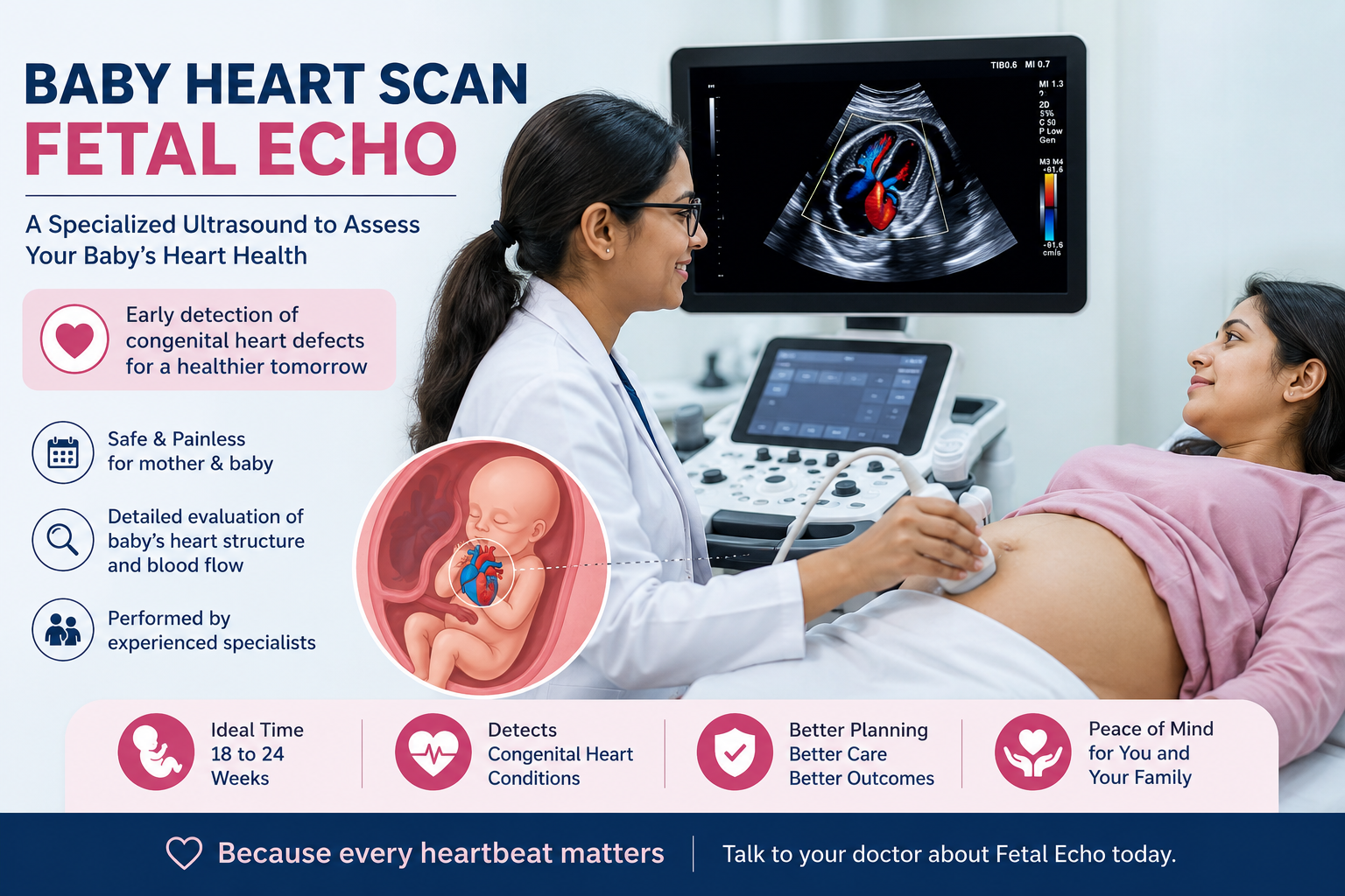
A fetal echo in Surat is a detailed ultrasound of your unborn baby’s heart, usually done between 18 and 24 weeks of pregnancy. This safe, painless scan checks the heart’s chambers, valves, rhythm and blood flow. Doctors advise it when there is a higher chance of a heart problem, so care can be planned early.
Key Takeaways
- A fetal echo in Surat looks closely at your baby’s heart structure, rhythm and blood flow before birth.
- Most scans happen between 18 and 24 weeks, though your doctor may suggest an earlier or repeat scan.
- The test uses sound waves only, so it is safe for both mother and baby.
- It is usually advised when family history, maternal health or a routine scan raises a concern.
- Early findings help your team plan delivery and newborn care with a heart specialist.
Table of Contents
- What is a fetal echo?
- Why would I need a fetal echo in Surat?
- When is the scan done during pregnancy?
- How is a fetal echo performed?
- What can a fetal echo detect?
- What happens after the scan?
- Frequently Asked Questions
What is a fetal echo?
A fetal echo, or fetal echocardiography, is a specialised ultrasound that studies your baby’s heart while it is still developing. Unlike a routine pregnancy scan, it focuses only on the heart. The test maps each chamber, valve and major blood vessel, and it records how the heart beats.
During the scan, a fetal medicine expert watches blood move through the heart in real time. Colour Doppler adds moving colours to show the direction and speed of that flow. As a result, even small differences in the heart’s build or rhythm become visible early.
Our fetal medicine specialist in Surat uses this scan to answer one key question. Is your baby’s heart forming and working the way it should? Because the heart is complex, this focused view offers far more detail than a standard anomaly scan alone.
Why would I need a fetal echo in Surat?
You may need a fetal echo in Surat when something raises the chance of a heart difference in your baby. Common reasons include a family history of heart defects, certain maternal conditions, or an unclear finding on your routine scan. Your obstetrician then refers you for this closer look.
Several situations make doctors recommend the scan. Consider the most frequent ones below.
- A parent or sibling was born with a congenital heart defect.
- The mother has diabetes, lupus, or another long term condition.
- Certain medicines or infections occurred early in pregnancy.
- A routine anomaly scan showed something that needs a second look.
- The pregnancy involves twins, IVF, or a raised nuchal translucency.
Even without these factors, some parents simply want extra reassurance. In that case, our team is glad to explain whether the scan adds value for you. If you are unsure, our fertility and pregnancy care team can guide the decision during your visit.
When is the scan done during pregnancy?
A fetal echo is usually performed between 18 and 24 weeks of pregnancy, when the heart is large enough to study in detail. Sometimes a specialist offers an early scan around 13 to 16 weeks, and a repeat scan later if a finding needs to be tracked over time.
The timing matters because the heart keeps growing and changing. The table below shows how the scan fits into the pregnancy timeline.
| Stage | Approximate weeks | Purpose |
|---|---|---|
| Early fetal echo | 13 to 16 weeks | Used for high risk pregnancies when an early look is helpful |
| Standard fetal echo | 18 to 24 weeks | Best window for a full, detailed heart assessment |
| Follow up scan | After 24 weeks | Tracks a known finding or checks the rhythm again |
Your exact schedule depends on your history and earlier results. Therefore, the specialist will set a plan that suits your pregnancy rather than a fixed date.
How is a fetal echo performed?
A fetal echo is done much like a normal ultrasound and takes about 45 to 90 minutes. You lie down comfortably, a warm gel is applied to your abdomen, and the sonologist moves a probe to capture the heart from many angles. The scan is painless and needs no injections.
Throughout the scan, the specialist studies the heart carefully. First, they check the four chambers and the valves between them. Next, they trace the large vessels that carry blood to and from the heart. Finally, colour Doppler confirms that blood flows in the right direction at a healthy speed.
Occasionally the baby’s position hides part of the heart. When that happens, the sonologist may ask you to walk for a few minutes or return later. This patience helps capture every view clearly, which keeps the report accurate.
What can a fetal echo detect?
A fetal echo can detect many congenital heart defects, rhythm problems, and issues with the valves or major vessels before birth. It cannot promise a perfectly healthy heart, yet it finds most significant structural differences early, which gives your family time to prepare and plan the right care.
The scan commonly looks for the following.
- Holes between the heart chambers, such as septal defects.
- Narrow or leaking valves that affect blood flow.
- Problems with how the large arteries connect to the heart.
- An irregular heartbeat that is too fast, too slow or uneven.
According to the American Heart Association, a fetal echocardiogram gives a detailed picture of the baby’s heart structure and function. When a difference is found, early knowledge allows a paediatric heart team to be ready at delivery. Outcomes vary from case to case, so your specialist will explain what any finding means for you.
What happens after the scan?
After a fetal echo, the specialist explains the findings the same day in most cases. If the heart looks normal, you simply continue your regular pregnancy care. If a difference is seen, your team plans further monitoring, counselling and a safe delivery with newborn heart support ready.
A calm, clear discussion follows every scan. The specialist describes what was seen, answers your questions, and outlines the next steps in plain language. Where needed, they coordinate with paediatric cardiologists so your baby has expert care from the first breath.
To book this scan or to understand your reports, you can reach our team through the appointment page or the contact page. Early guidance brings peace of mind, and it helps you focus on a healthy, hopeful pregnancy.
Frequently Asked Questions
Is a fetal echo safe for my baby?
Yes. A fetal echo uses sound waves only, not radiation. Decades of use show it is safe for both mother and baby. The scan is painless, needs no dye or injection, and can be repeated when your specialist feels it is helpful.
How long does a fetal echo take?
Most fetal echo scans take about 45 to 90 minutes. The exact time depends on your baby’s position and how easily the heart can be seen. If the view is unclear, the sonologist may pause and try again for accurate images.
Will a fetal echo find every heart problem?
A fetal echo finds most major heart differences, but not every tiny one. Some issues appear only after birth. Even so, the scan detects the conditions that matter most for planning delivery and early newborn care, which improves preparedness.
Do I need a full bladder for the scan?
Usually no. Unlike some early pregnancy scans, a fetal echo in the mid trimester rarely needs a full bladder. Your clinic will share any simple instructions beforehand, so you can arrive relaxed and comfortable for the appointment.
Who performs a fetal echo in Surat?
A trained fetal medicine specialist or sonologist performs the scan, often alongside a cardiologist for complex cases. At Paaranu IVF in Surat, an experienced team reviews your history first, then tailors the scan and the report to your pregnancy.
What should I do if a problem is found?
Stay calm and lean on your care team. Many heart differences can be managed with a clear plan, monitoring and timely newborn care. Your specialist will explain the finding, arrange further tests if needed, and support you through each step.
Book a consultation at Paaranu IVF Clinic in Surat to schedule your fetal echo or to review your scan reports. Call +91 98797 72259 or email care@paaranuivf.com, and our fetal medicine team will guide you with warmth and clarity.
About Paaranu IVF
Paaranu IVF, part of Paaranu Women’s Superspecialities, is a dedicated fertility and women’s health centre in Surat, Gujarat. The clinic offers IVF, IUI, ICSI, male infertility care, fetal medicine and advanced gynaecology under one roof. Families across Surat and South Gujarat trust Paaranu IVF for calm, expert and personalised care on their journey to parenthood.